
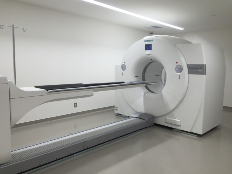
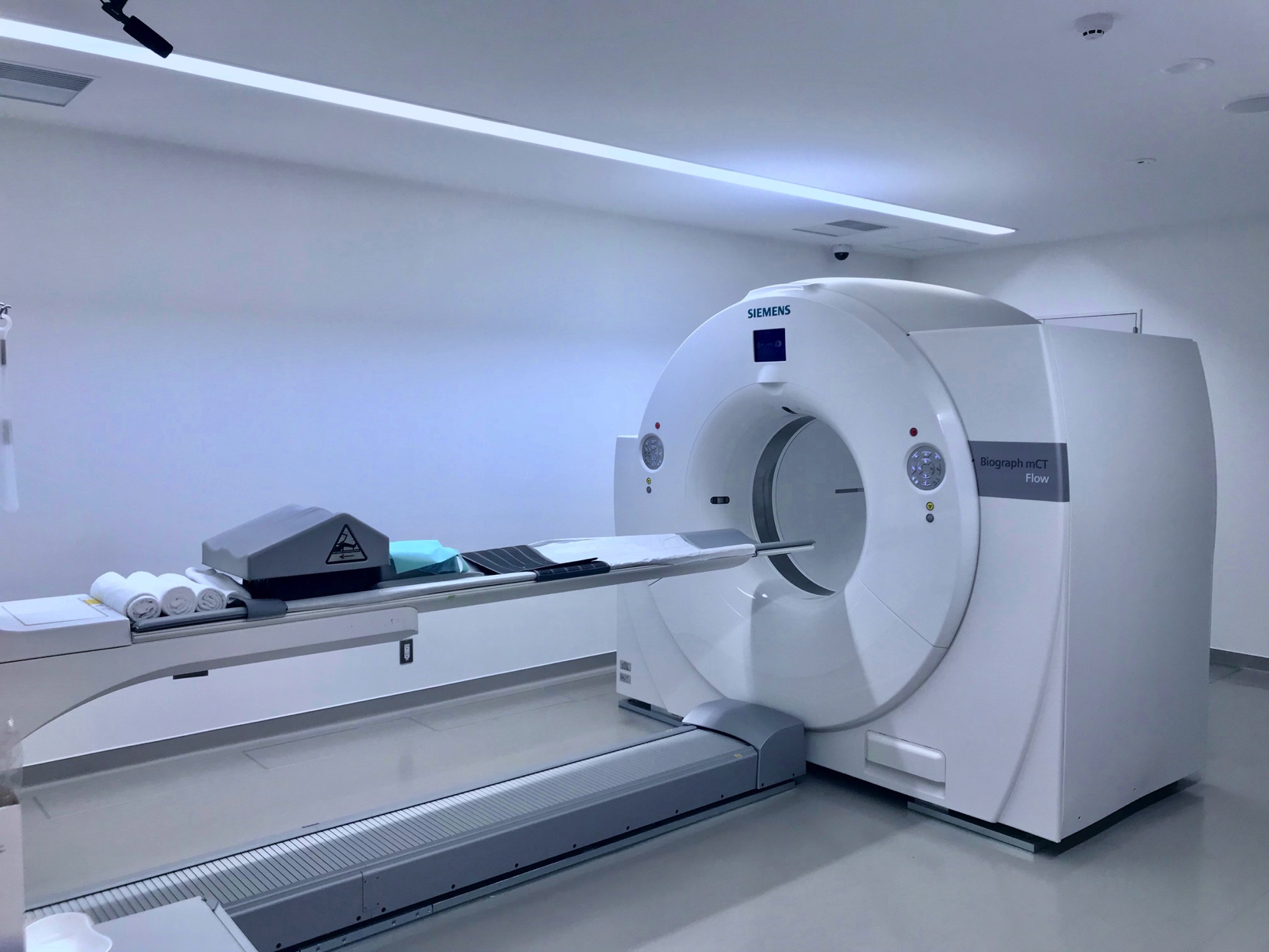
 Biograph mCT Flow (SIEMENS)
PET(Positron Emission Tomography) is an imaging technique which observes the distribution of positrons in the body. Because cancer cells divide more frequently, they consume an extraordinary amount of glucose -- about three to eight times more than normal cells. PET diagnostics utilize this characteristic to examine cancer.
We inject FDG (a tracer with similar composition to glucose) into the patient’s body, and perform diagnosis by detecting FDG accumulation in the body. Conventional CT or MRI methods detect cancer by image. On the other hand, PET detects cancer through the characteristics of cancer cells. FDG accumulation is directly proportional to the amount of glucose that cancer cells consume, in other words, it is proportional to the activity of cancer cells. For this reason, we consider PET to be a method for detecting cancer by the function of the cancer cells themselves.

The gantry aperture of our PET/CT unit is 780 mm in diameter. The generous aperture size reduces the sensation of confinement, and even patients who are not comfortable in small spaces can undergo examination with less anxiety than conventional diagnostic units.
Furthermore, the bed can accept body weights of up to 227 kg, so even heavily-built patients may safely undergo examinations.
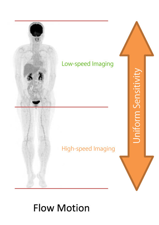
We use a PET image collection technique known as Flow Motion, which utilizes variable speed, continuously moving scanning. Flow Motion allows us to specify the imaging range as required, and change the imaging speed for each section.
Therefore, at the sections of interest, we can use lower speeds to gather clearer images, with improved SNR (Signal to Noise Ratio). Outside of the target area, we can use higher speeds to shorten the examination time. Furthermore, excellent uniformity of rostrocaudal sensitivity allows Flow Motion to collect images of uniform quality from the head to the lower limbs.
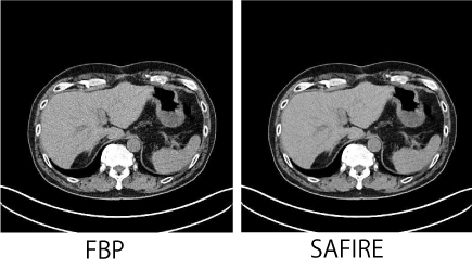
The SAFIRE hybrid iterative reconstruction method may be utilized to reduce exposure to radiation.
When we perform CT image reconstruction, SAFIRE makes it possible to record images at lower X-ray doses. Compared to conventional FBP (Filtered Black Projection) methods, this results in an approximately 50% decrease in radiation.
 Examination Room
 Hall
Hall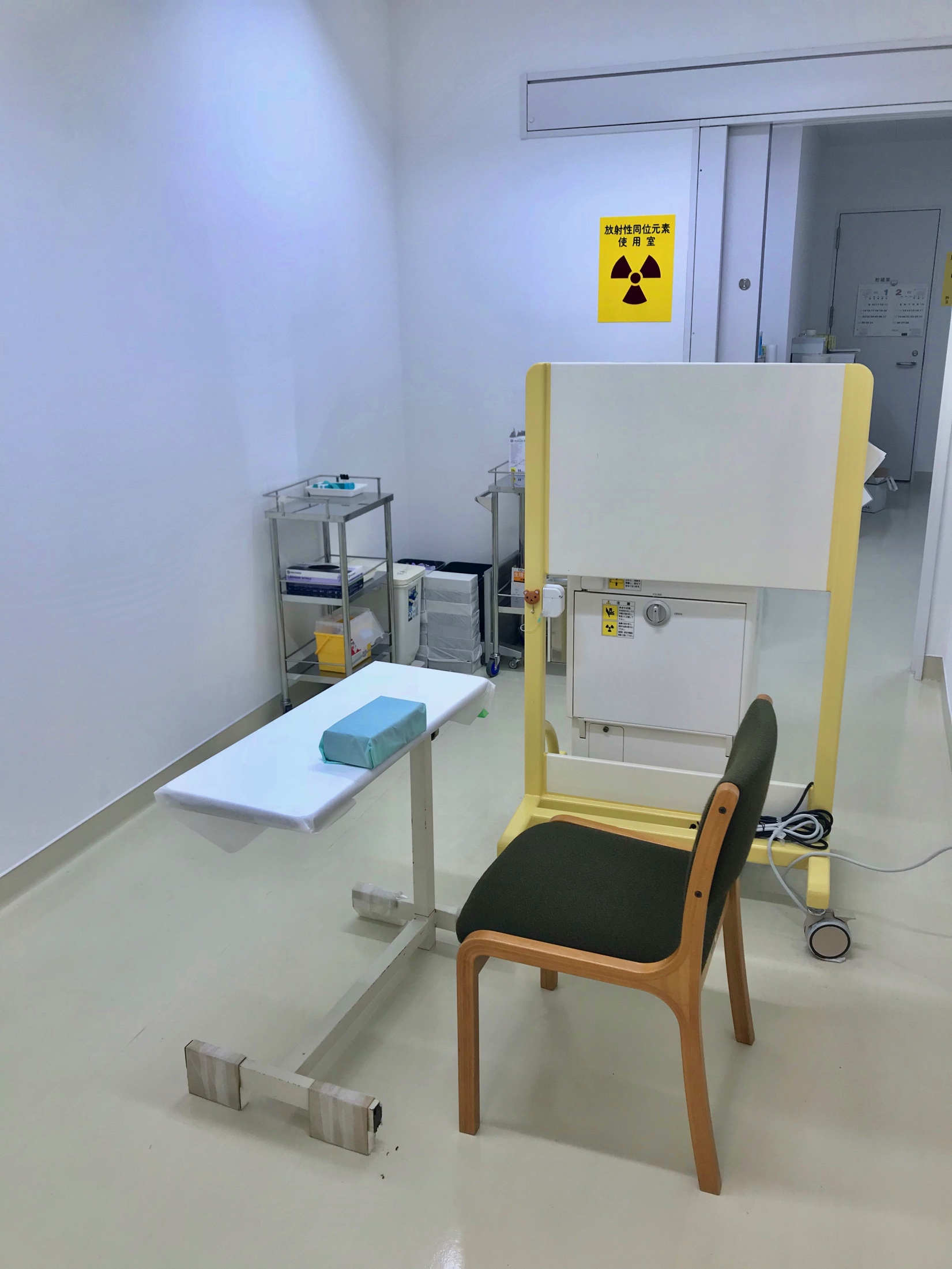 Treatment Room
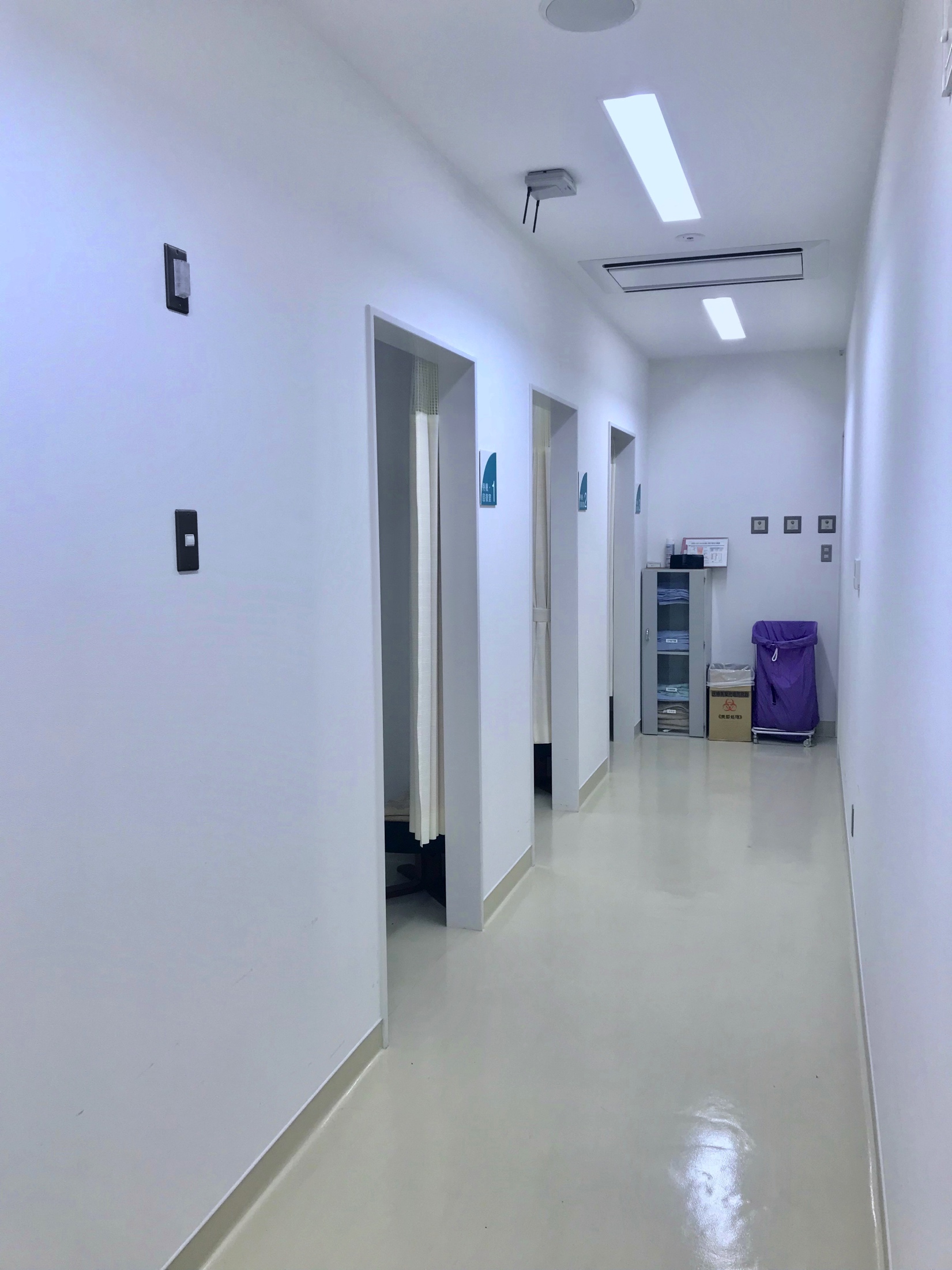 Waiting Room
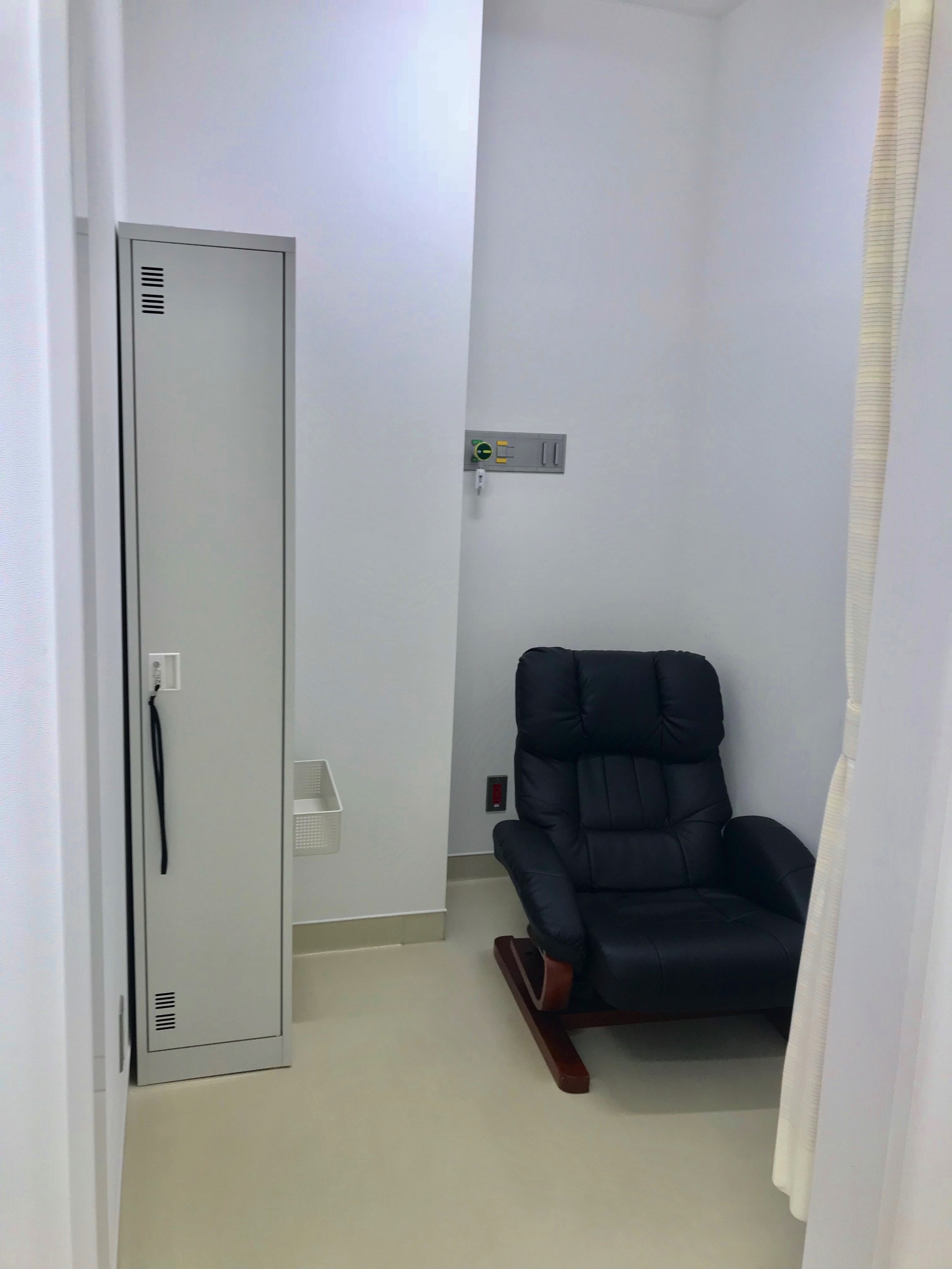 Waiting Room
○First-Time Patients / Returning Patients (without appointment)
8:30~11:00
○Automated Reception for Returning Patients (Reservation Only)
8:00~17:00
First-time patients are expected to bring a letter of introduction. For more information, please refer to the Outpatient Guide